Повышенная экспрессия генов AKR1C2 и AKR1B1 связана с развитием резистентности к доцетакселу при раке предстательной железы.
Повышенная экспрессия генов AKR1C2 и AKR1B1 связана с развитием резистентности к доцетакселу при раке предстательной железы.

Аннотация
Лекарственная устойчивость является одной из наиболее актуальных проблем в современной онкологии и представляет собой результат сочетания различных молекулярных событий в опухолевых клетках под действием препарата, полное понимание которых еще предстоит определить. Настоящее исследование было сосредоточено преимущественно на оценке экспрессии генов семейства альдо-кето редуктаз при развитии химиорезистентности в случае рака предстательной железы (РПЖ) – одного из наиболее распространенных и социально значимых видов рака у мужчин во всем мире. С помощью транскриптомного профилирования клеточной модели РПЖ в процессе развития резистентности к таксану доцетакселу, было показано статистически значимое повышение экспрессии двух генов из семейства альдо-кето редуктаз – AKR1C2 и AKR1B1, что также было подтверждено методом количественной ПЦР. Полученные результаты подчеркивают высокий потенциал этих генов для дальнейших исследований в качестве перспективных маркеров резистентности, а также терапевтических мишеней.
1. Introduction
Prostate cancer (PC) is a highly prevalent and socially impactful malignancy that affects males globally. In Russia, the incidence of PC is also notably high, with 40,785 newly reported cases in 2017 . Androgen deprivation therapy is widely recognized as the leading strategy for managing PC. However, despite extensive research and advanced treatment, patients invariably develop castration-resistant PC (crPC), a highly aggressive and lethal form . The initial treatment choice for crPC patients typically involves chemotherapy utilizing taxanes, which are cytostatic drugs that bind to beta-tubulin in microtubules, disrupting critical cellular processes . The significant challenge in oncourology lies in the inevitable development of chemoresistance to taxane-based treatments. Aldo-ketoreductases (AkRs) are a class of oxidoreductases primarily responsible for converting carbonyl groups into alcohols through a reduction mechanism . In humans, fifteen different AkRs have been discovered, and while the particular carbonyl substrates they act upon may differ, these proteins possess specific structural traits in common. The upregulation of AkRs has been observed in diverse tumor categories, including PC , , . Furthermore, scientists have proposed their potential role as tumor biomarkers, indicating the significance they hold. Moreover, multiple studies have established a link between the expression of AkRs genes and the emergence of resistance to several prominent chemotherapy drugs . Our study focused on addressing the core inquiries concerning the molecular mechanisms underlying tumor resistance to various drugs. Specifically, our aim was to investigate the variations in the expression of AkRs genes during the process of resistance development to docetaxel (DoX) using an experimental model of PC cells.
2. Research methods and principles
PC3 cells were cultured in RPMI-1640 medium at a temperature of 37C with a 5% CO2. To generate resistant cell sublines, we employed a gradual increase in DoX concentration in the medium. Cells that survived at a DoX concentration of 10 nM were deemed resistant. We performed RNA extraction using the RNeasy Micro Kit (Qiagen) and library preparation using the KAPA RNA HyperPrep Kit with RiboErase (Roche). Next-generation sequencing (NGS) was conducted on the NextSeq 2000 instrument. Analysis of differential gene expression was carried out utilizing the edgeR in the R. To perform reverse transcription, we utilized the Mint kit (Evrogen) and conducted quantitative polymerase chain reaction (qPCR) on the Applied Biosystems 7500 instrument with three technical replicates. The HPRT1 gene was used for normalization. The relative mRNA expression level for each comparison was determined using the dCT method. Statistical significance of gene expression differences was assessed with a criterion of p<0,05.
3. Main results
The key findings related to the varying expression of AkRs genes in different experimental cell groups during DoX treatment are summarized in Table 1. When comparing the DoX 4 nM and 10 nM groups, the results showed statistically significant findings for the AKR1C2 and AKR1B10 genes. Importantly, the differential expression of AKR1C2 and AKR1B10 exhibited a remarkable increase in DoX resistant cells compared to cells treated with DoX initially. Specifically, AKR1C2 expression showed a 195-fold increase, while AKR1B10 expression showed a 10.63-fold increase. Furthermore, in the comparison between resistant cells and the control group, the expression of AKR1C2 was enhanced by a 3.2-fold increase, while AKR1B10 expression showed a 2.19-fold increase. Additionally, when comparing the DoX 8nM and DoX 10nM experimental cell groups, there was a notable and statistically significant upregulation of AKR1B10 and AKR1C2 expression. Notably, the differential expression of AKR1C2 showed a remarkable 10-fold increase in this comparison.
Summary results of AkRs genes differential expression across different experimental cell groups
Log2FC – fold change in expression, binary logarithm; FDR - false discovery rate; 1- Control vs. DoX 10nM, 2 - 4nM vs. DoX 10nM, 3-8nM vs. DoX 10nM
Gene | Log2FC1 | FDR1 | Log2FC2 | FDR2 | Log2FC3 | FDR3 |
AKR7A3 | 0,84 | 1,0E+00 | 0,57 | 1,0E+00 | 0,81 | 1,0E+00 |
AKR1A1 | 0,05 | 1,0E+00 | 0,18 | 8,0E-01 | 0,35 | 4,7E-02 |
AKR7A2 | 0,28 | 8,2E-01 | 0,51 | 3,2E-02 | 0,85 | 3,0E-05 |
AKR1B1 | 1,09 | 7,5E-02 | 2,26 | 7,0E-05 | 1,67 | 1,7E-03 |
AKR1B10 | 1,13 | 3,3E-02 | 3,41 | 2,1E-08 | 1,66 | 6,5E-04 |
AKR1C3 | 0,3 | 1,0E+00 | 2,2 | 7,9E-06 | 0,9 | 8,5E-02 |
AKR1E2 | 0,66 | 1,0E+00 | 1,09 | 7,7E-01 | 0,7 | 1,0E+00 |
AKR1C2 | 1,67 | 3,1E-02 | 7,61 | 1,5E-07 | 3,34 | 5,4E-05 |
AKR1C1 | 1,03 | 5,2E-02 | 2,51 | 1,5E-06 | 1,07 | 3,0E-02 |
The evaluation focused on the changes in gene expression levels of AKR1C2 and AKR1B10 after being exposed to DoX treatment. The findings demonstrated a significant rise in AKR1C2 expression within the DoX 10 nM group, as compared to both the DoX 4 nM group (p = 0.001) and the DoX 8 nM group (p = 0.002) (Fig. 1). In particular, the AKR1C2 expression in the DoX 10nM group showed a significant 13.82-fold rise compared to the DoX 4nM group. Moreover, there was a notable 4.88-fold increase in AKR1C2 expression in the DoX 10nM group in comparison to the DoX 8nM group.
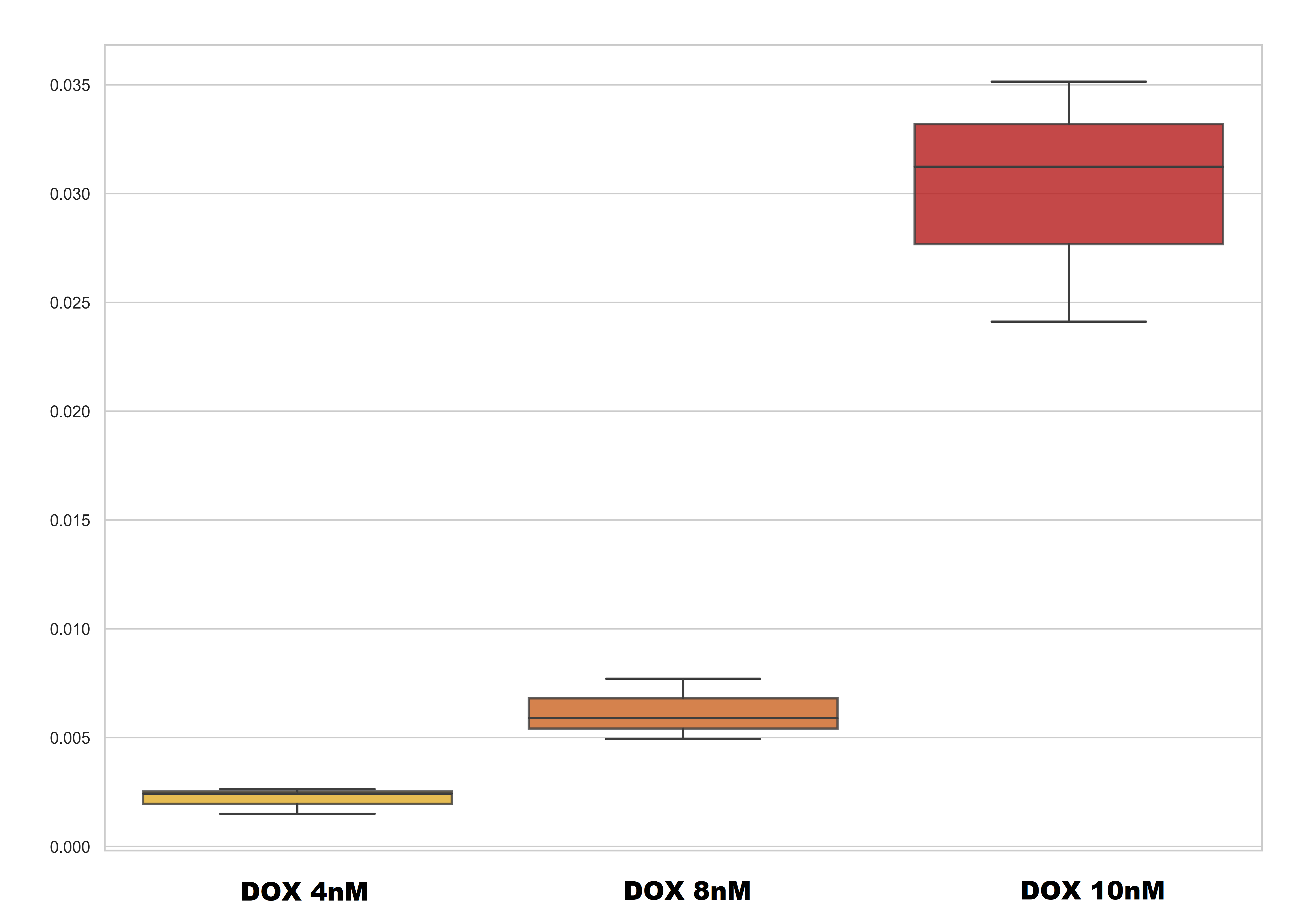
Boxplots of relative expression of AKR1C2 mRNA levels among different experimental groups
the y-axis represents the 2-ΔCT values
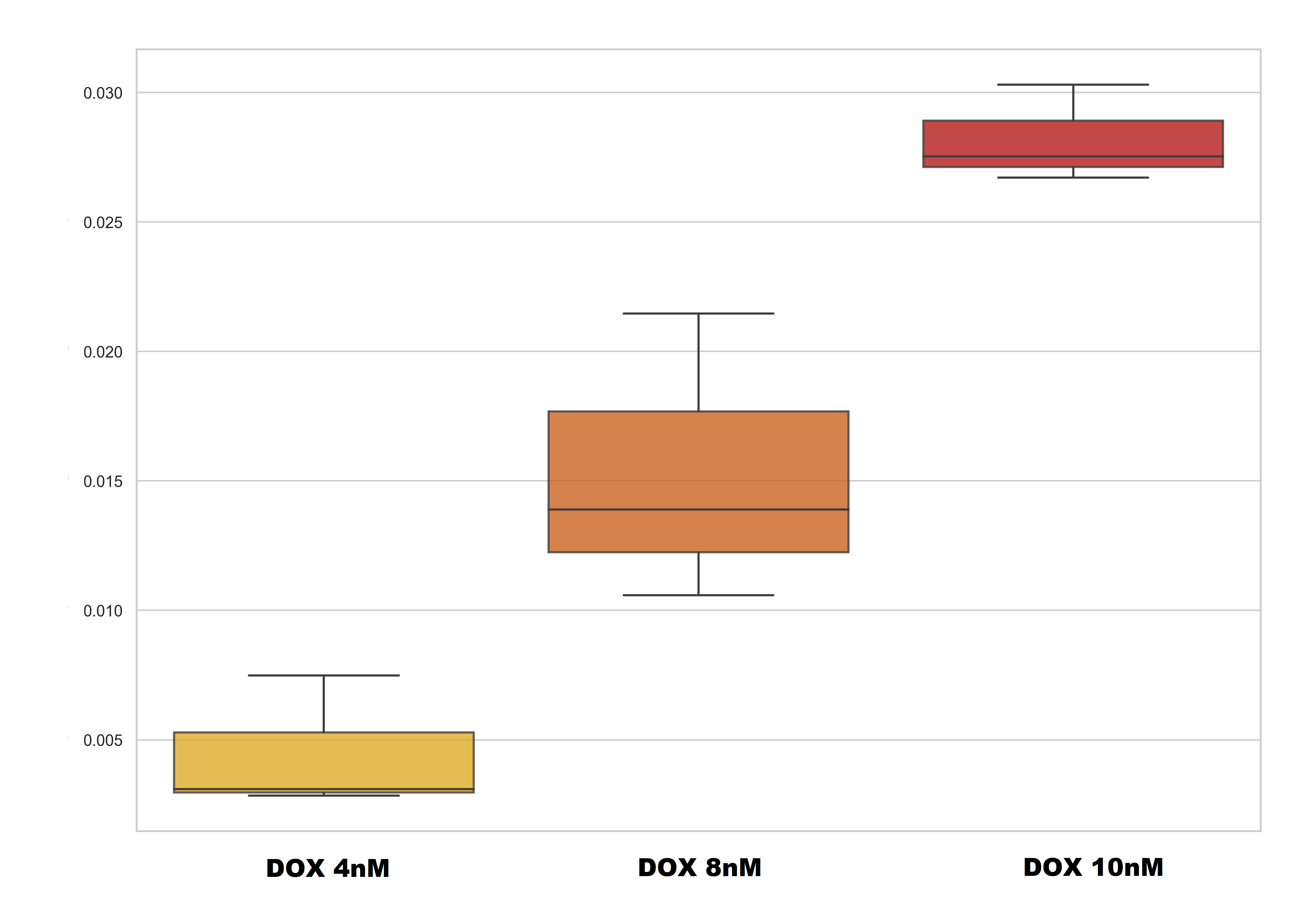
Boxplots of relative expression of AKR1B10 mRNA levels among different experimental groups of cells
the y-axis represents the 2-ΔCT values
4. Discussion
Reviewing the literature, it has been observed that the overexpression of the AKR1B10 gene is frequently found in different tumor types and may be linked tothe disease . Data from multiple studies have revealed connections between elevated expression of this gene and the emergence of chemoresistance to therapeutic agents across various cancer types , . Numerous studies have discovered a correlation between heightened AKR1C2 gene expression and tumor resistance to diverse chemotherapeutic treatments in glioblastoma, lung cancer, and breast cancer , , , . Additionally, the AKR1C2 gene plays a role in regulating steroid hormone ligands and receptors, potentially linking it to the advancement of hormone-dependent cancers like breast cancer and PC . The findings from this study align with previous literature and highlight the potential significance of the AKR1C2 and AKR1B10 genes in the chemotherapy resistance to DoX in crPC. The complete understanding of how these genes interact with each other is still unknown.
5. Conclusion
Chemotherapy resistance continues to be a major obstacle in the clinical use of these drugs. Uncovering potential therapeutic targets involved in resistance could greatly enhance the efficacy of these agents and ultimately enhance patient survival rates. In the study conducted on PC cells as a model, the researchers examined the differential gene expression of AkRs during the development of resistance to the taxane drug, DoX. The expression levels of these genes have been found to be higher in different tumor types, suggesting their potential involvement in chemoresistance. The study results indicate a strong statistical correlation between the overexpression of two specific genes, AKR1C2 and AKR1B10, and the development of resistance to DoX in PC3 cells.