МЕДИЦИНСКАЯ ДИАГНОСТИКА НА ОСНОВЕ МИКРОСТРУКТУРНОГО АНАЛИЗА БИОЛОГИЧЕСКИХ ЖИДКОСТЕЙ КАК ЗАДАЧА БИОИНФОРМАТИКИ

MEDICAL DIAGNOSTICS BY MICROSTRUCTURAL ANALYSIS OF BIOLOGICAL LIQUID DRIED PATTERNS AS A PROBLEM OF BIOINFORMATICS
Funding
This work has been supported by the Russian Foundation for Basic Research, project no. 15-03-08050
Conflict of Interest
None declared.
Peter Lebedev-Stepanov1,2,*, Marina Buzoverya3 , Irina Shishpor3 , Konstantin Vlasov3 , Yulia Potekhina4
1Photochemistry Center FSRC “Crystallography Photonics” RAS, Moscow, Russia,
2National Research Nuclear University MEPhI, Moscow, Russia,
3Sarov State Physics Technical Institute, Sarov, Russia,
4State Medical Academy, Nizhny Novgorod, Russia
*To whom correspondence should be addressed.
Associate editor: Giancarlo Castellano
Received on 03 November 2017, revised on 04 December 2017, accepted on 11 December 2017.
Abstract
Motivation: It is important to develop high-precision computerized methods for quick medical diagnostics based on the unique clinical experience obtained within the past decade. This method provides specialized solutions for diagnostics and the monitoring of specific diseases, identifies reserves of human health, and takes actions to prevent the depletion of these reserves. In this paper we present one of the new directions in bioinformatics, i.e. medical diagnostics by means of an automated expert system on the basis of morphology analysis of digital images of biological liquid dried pattern.
Results: The proposed method is a combination of bioinformatics and biochemistry approaches for obtaining diagnostic information from a morphological analysis of standardized dried patterns of biological liquid sessile drop. We conducted our own research in collaboration with medical diagnostic centers and formed an electronic database for the recognition of the following types of diseases: neoplasms; diabetes mellitus; diseases of the circulatory system; cerebrovascular disease; diseases of the digestive system; diseases of the genitourinary system; infectious diseases; factors relevant to the work; factors associated with environmental pollution; factors related to lifestyle. The laboratory instrument for diagnostics of the human body in pathology states has developed and the diagnostic results have been analyzed.
Availability: Access to the testing of software can be obtained upon request via the contact email below.
Keywords: dried pattern, microstructural bio-liquids’ analysis, quantitative microscopy, cracks, concretions, microfluidics, automatic expert system, descriptors, bioinformatics.
Contact: petrls@yandex.ru
1 Introduction
In current clinical practice, there are a variety of methods for diagnostics of the human health. There is an active search for the improvement of diagnostic techniques when determining the objectives of body response to the impact of external factors, including radiation, radioactivity; to determine the natural resistance and adaptation reserves of the body, to predict the direction of the pathological process (especially the diagnosis of cancer), to accelerate the diagnostic analysis with high accuracy while minimizing cost consumption. It is important to develop high-precision computerized methods for medical rapid diagnostics, which are generalizing the unique clinical experience obtained in the past decade as specialized solutions for diagnostic problems for the control of specific diseases and, potentially, for wider health monitoring, identifying the reserves of human health and the prevention of these reserves from being depleted. On the one hand, the elaboration of complex and mass rapid diagnosis is the creation of multi-function, multi-parameter stationary systems and, on the other hand, in the field of mobile diagnostic devices, through which the patients can receive information about their health which can in turn be forwarded to the diagnostic center by Internet or telephone network. Despite the fact that the list of diagnostic techniques is a comprehensive assessment of the patient's condition and can contain hundreds of names, the widespread use of such diagnostic practices is limited by their material and time expenses. Thus, the desired result is the development of a method that combines the simplicity and speed of sample preparation with the versatility and reliability of diagnostic information, with a view to taking into account the individual characteristics of each patient. Also, mobile or semi-mobile diagnostic technologies are working individually with each patient not only with the state of disease but also in the phase of preventive diagnosis when symptoms are still not fully clear. Such technologies make the patient an active participant in diagnostics (Topol, 2012). Diagnostic methods based on the analysis of biological liquid drying spot (pattern) have all these advantages. Applications and techniques of dried blood spot (DBS) sampling (DBS-based polymerase chain reaction technologies, LC-MS/MS methods for quantitative analysis of small molecules, digital microfluidics platform for DBS diagnostics, etc.) have been reviewed in (Li et al, 2014). In addition, to date there has been the accumulation of a large amount of actual data on specific features of the structure formation of different bio-liquids of the human body in the normal state and during the pathological state development. A study of mechanisms for bio-liquids’ structure formation by a method of optical microscopy is of specific interest (Shabalin et al, 2001; Buzoverya et al. 2012; Lebedev-Stepanov et al, 2013, Brutin, 2015). Dried pattern sample is formed by the evaporation of a small drop of the biological liquid on a flat substrate. Such a pattern can be used for the diagnosis of various diseases. There is only a small number of unique medical experts in the world who, based on their own many years of experience in practical research, can exactly determine the presence of the pathology by studies of pattern structure. The widespread implementation of this method is limited to a small number of professionals with the required qualifications. However, high informational content of pattern structure and the uniqueness of the correspondence between the deviations of pattern structure with pathology from the normal state make it possible to develop the science-based, highselective diagnostic method based on standard computerized analysis procedures integrated with unique and expert experience. Thus, it could be possible to create software and hardware complexes for users with low medical qualifications. Moreover, such a diagnostic system allows the development of a standard methodology for recognizing the specific nature of the deviation from the normal structure of the pattern, which is yet not included in the list of diseases defined above by unique experts and without their involvement. The problem is the creation of a hardware and software system that implements the principle of diagnosis based on the standard procedure of pattern preparation, including digital recognition of images and its computer analysis based on specially developed algorithms by comparing with expert database descriptors.
2 Methods
The method is based on the conversion of bio-liquids into a solid phase through dehydration on a substrate and the study of digitized images of thin film pattern obtained from a computerized expert system. When dehydrating a bio-liquid by osmotic forces and other physical-chemical processes, molecules and molecular complexes are distributed in strictly defined locations over the drying drop area in the form of concentration zones. As a result, the film is formed, possessing a structure, whose specificity is defined by chemical composition and character of that presented in bioliquid substance interaction. Films’ appearance is applied for the diagnostics of a wide range of diseases. Pathological changes, occurring in the body, lead to violation of qualitative and the quantitative composition of bio-liquids, which influence the solid phase morphology. As a result of earlier conducted studies, there was the development of a specific approach to the analysis of dehydrated biologic liquids, which permitted the obtainment of new data on the human health state; to reveal diseases at early stages, as well as to classify them by the form and severity of pathology. It was shown that the performance of systematic studies of biologic liquids within the frames of the chosen single approach contributed to the development of methods of microstructural bio-liquids analysis and to the understanding of observed bio-structural effects in drying drops of multi-component liquids (Buzoverya et al. 2012). Samples were prepared by drop dehydration on open solid substrate (microscope slide). Drops of prepared solution (volume of 0.01 - 0.02 ml) were deposited on the substrate surface by a microdispenser. This sample is dried at a temperature from 20 to 25 degrees Celsius and with a relative humidity of 28 to 35 % minimal mobility of ambient air. The duration of the drying phase is from 12 to 24 hours. The result is a thin dried pattern which is suitable for further analysis. Note, that the diameter of the pattern on the substrate should not exceed 5-7 mm of dried pattern and completely obstructing the field of view of the microscope. To obtain the digitized image of the dried pattern, the microscope Polam R211-M (LOMO, Russia) was used (Figure 1).
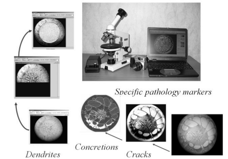
Fig. 1 - Special components of program library allow to digitize of structural elements of dried pattern and calculate the pathology markers parameters (a number and sizes of objects, their distribution uniformity, arrangement symmetry, deviation angle, fractality et al.).
To obtain quantitative characteristics and further application of mathematical apparatus, the development of computer systems for image processing is required. Presently, computer microscopic systems provide wide capabilities for a many-sided study of microscopic image structures. The setup is formed by microscope, web-camera and computer. In the majority of cases these systems provide a set of standard operations: computer image loading, a change of morphologic and brightness characteristics of studied objects, and a number of service operations. A hardware-software complex (experimental setup) for dried pattern image processing has been developed. The complex contains a program library that makes it possible to process images with a different biochemical component (blood serum, gall, tear et al.), to assist in distinguishing among structural elements, to reveal contours and to measure the relationship among areas of structural objects of dehydrated bio-liquid. Special programs for digitizing and quantitative analysis of specific pathology markers in the bioliquid structure have been developed. The complex represents a research computer system for routine analysis in medical and biological applications and fundamental studies in the field of functional bio-liquids morphology (Buzoverya 2013). The complex permits the processing of images for any body’s bio-liquids, biopolymer and polymer films and for the acquisition of quantitative structure characteristics. Software implements the previous experience in methodology of studying a structure of polymeric systems and other solid body objects. It is intended for broad use in the field of investigating different structures of multicomponent systems, including biopolymer films, and involves: - programs ProtoBlood, Concretion, which allow the digitizing of structural dried pattern elements and for the calculation of most image parameters (a number and sizes of objects, their distribution uniformity, arrangement symmetry, deviation angle, fractality et al.); - a program Saliva which measures the relationship among areas of structural objects of dendrite structure dried pattern, conducts recognition and dispersion analysis of dendrites, measures and calculates a large number of image parameters (dried pattern concentration zone areas, sizes and the number of dendrites, a degree of their brunching, a distribution character et al.); - a program for microstructural defects’ analysis MarkerBlood, which allows the calculation of the number of specific objects and the total area occupied by them; - a program for quantitative microscopy of object images with dendrite structure Dendrit – calculates the total area of different concentration zones and evaluates their color, calculates the number of specific objects; - programs Calibration, Fractals for calibration and fractal analysis. The program library is being constantly enlarged through development of new software, oriented at both fundamental and prompt routine measurements. The main methodological tasks for the performance of quantitative microscopy in clinical studies, is the specification of structures’ characteristics which are most informative when viewed from the perspective of the medicine problem. For example, in dried pattern with a protein component (blood serum, gall, and liquor et al.) the main structural elements are cracks and concretion. Dried pattern images also illustrate different characteristics of the structure formation of human blood serum, of the human body in the normal state and when the pathological process is being developed.
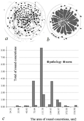
Fig. 2 - Round concretions in blood serum patterns: a – pathology; b –norm; c – distribution of concretions by size.
Cracks and concretions form in the process of bio-liquid dehydration. Due to a complex component composition this process occurs phase-by-phase accompanied by the formation of solid phase concentration zones, which are formed through corresponding bio-liquid components with definite physicochemical parameters. After the evaporation of free water, a bio-liquid drop fully goes over into a solid phase and forms a dried pattern. As a result of bound water evaporation being continued, in the dried pattern structure there develops rather powerful processes of stretching and the compression of material; also due to protein molecules’ coagulation, which causes dried pattern cracking and structural element formation – cracks. Concretions are formed as a result of homogeneous substance accumulation in different dried pattern zones, as a rule, surrounded by cracks. In the course of experimental and clinical studies it has been shown that cracks and concretions possess a well-defined interrelation with definite physiological and pathological states of the human body. Therefore, the study of quantitative characteristics of cracks and concretions (a form, distribution homogeneity over the dried pattern area, size, number and et al.) provides information on the microstructural dried pattern inhomogeneity. To illustrate the program ProtoBlood operation on digitizing the cracks, let us give an example of blood serum dried pattern image processing in two independent samples: practically healthy humans – the norm (42 dried patterns samples) and patients with the Cerebral atherosclerosis – the pathology (36 samples). Typical samples for each of these samples are given in Figure 2, these pattern images digitized by cracks – in Figure 2 a,b. Figure 2c presents a histogram of distribution of correct round shape concretions by size. The histogram shows that the dried pattern of healthy human blood plasma has large concretions of rounded and oblong forms, located mainly radially. If the concretions are small, irregularly shaped, located chaotically, then there is a violation of the lipid balance of the blood plasma. Such violation indicates a patient with ischemic heart disease (Poteknina 2004).
3 Discussion
The device application has allowed the obtainment of interesting results, not only in biomedical applications, but also the acquisition of interesting data on the processes of dried pattern structure formation (self-organization of the multicomponent colloidal fluids and solutions). It is obvious that the further improvement of this method is an interdisciplinary R&D problem including medical, engineering, physical, and mathematical aspects. From a medical point of view, we have to legitimatize and justify the connection between the specific change in the pattern structure from a normal one, and the presence of a corresponding pathology; true selection of diagnostic descriptors. Taking into account mathematical and computation points of view, it is about creating an optimal algorithm of computer processing for the realization of the principle "from abnormal pattern structure to specific diagnosis of pathology" based on the proposed structural descriptors, maximizing the percentage of recognition for pathology; the creation of an optimal software package. From an engineering point of view, the problem is to provide the optimum instrument system including a sample preparation system, the computer to digitize the image, analysis and diagnostic information issue. From the point of view of physics, it is an elaboration of a physical model with an aim at optimally interpreting the structural features of the dried pattern and justifying the descriptors set for diagnostic interpretation. We are also elaborating on the useful software complex to predictive modeling of setup, spreading, evaporation of liquid droplet of inkjet size, as well as self-assembly of solvated monodisperse nanoparticles from the drop during evaporation. The most difficult case for modeling is a drop of biological liquid (such as blood serum, tear, saliva et al.) which consists of many different components of solution and forms the complex dry pattern onto substrate as a final stage of the solvent evaporation process. Some tasks should be especially emphasized: 1) Obtaining new experimental data for the design and development of adequate models. 2) The development of 3D models describing the redistribution of the components, the movement of the front phase and the evolution of the profile of the drying colloidal droplets with salt admixtures. 3) The inclusion of the viscosity of a colloid with time and concentration of salts needs to be included in the model variations, as well as changes of the vapor flux above the free surface of the droplets when the phase boundary (sol-gel) is moving. (Tarasevich 2004; Tarasevich et al. 2016). Modern computer methods are not yet able to design and create such a system in detail. Our current investigations propose the further development and the continual generalization of dissipative particle dynamics methods to describing the formation of the dry pattern and search for new interpretations of this process and pattern diagnostics optimization. The development of physicochemical models of the process makes it possible to suggest new static and dynamic descriptors for computer recognition of pattern image.
Список литературы
Brutin D. (2015) Droplet Wetting and Evaporation. From Pure to Complex Fluids. Academic Press. 464 P.
Buzoverya M.E., Shishpor I.V., Potekhina Yu.P., Shcherbak Yu.P. (2012) The bio-liquids’ microstructural analysis. Technical Physics. 2012; 82;87; 123–128. doi: 10.1134/S1063784212070079.
Buzoverya M.E., Shishpor I.V., Sadovoy S.A. (2013) Quantitative microscopy of images of dendrite structure objects. Certification on official registration of computer program No2013610336 (RU).
Lebedev-Stepanov P.V., Vlasov K.O. (2013) Simula-tion of self-assembly in an evaporating droplet of colloi-dal solution by dissipative particle dynamics. Colloids and Surfaces A: Physicochem. Eng. Aspects. 432; 132–138. doi: 10.1016/j.colsurfa.2013.05.012.
Li W., Lee M.S. (2014) Dried Blood Spots: Applica-tions and Techniques. John Wiley & Sons. 364 P.
Potekhina Yu. P., Zubeeva G.N,, Motylev I.M., Buzoverya M.E. (2004) Issledovanie struktury degidrati-rovannoy plazmy krovi bol’nykh s ishemicheskoy boleznyu serdtsa [Investigation of the structure of dehy-drated blood plasma of patients with ischemic heart dis-ease] // Medicinskaya Fizika. 1; 46–48.
Shabalin V.N., Shatokhina S.N. (2001) Morfologiya biologicheskikh zhidkostey cheloveka. [Morphology of the biological liquids of the human]. Мoscow: Khrizostom.
Tarasevich Yu. Yu. (2004) Mechanisms and models of the dehydration self-organization in biological fluids// Physics-Uspekhi. 47; 717-728.
Tarasevich Yu.Yu., Tarafdar S., Dutta T. (2016) Pattern formation in desiccated sessile colloidal droplets with salt admixture: Short review// arXiv:1611.00882 [cond-mat.soft]
Topol E. (2012) How the Digital Revolution Will Cre-ate Better Health Care, Basic Books, 303 P.